|
The Treatment Plan is a 3 page document.
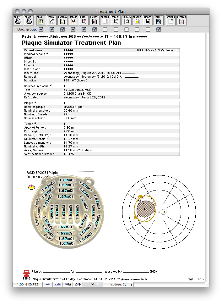
- Page 1 provides a table of patient identifiers, date & time of treatment, some radionuclide, plaque and tumor properties, a facial picture of the plaque and a miniature retinal diagram showing tumor location.
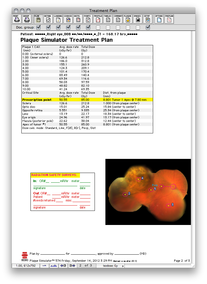
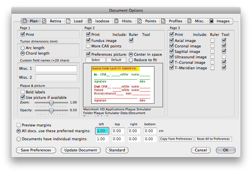
- On page 2 there is a table of point dose calculations along the central axis of the plaque (or tumor), at the prescription point, lens, macula, etc..., a thumbnails of the fundus image, and an optional picture. The default picture is a radiation safety survey form.
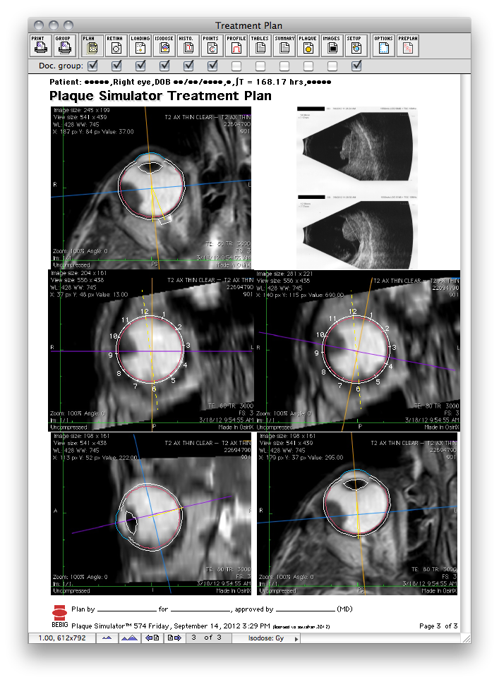
- Page 3 contains thumbnails of the CT or MR images used to model the eye and any ultrasound images used to measure or model the tumor dome.
- The page advance buttons along the bottom of the window change the page being previewed.
|