|
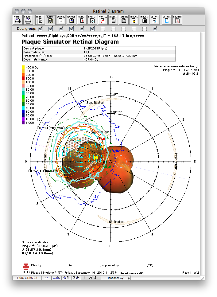
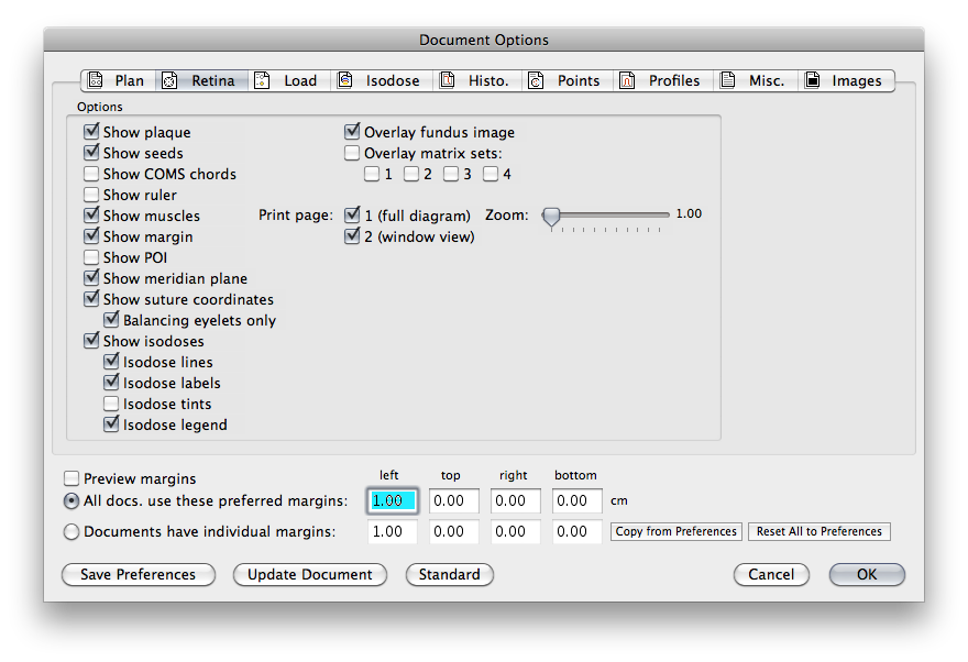
The Retinal Diagram can be a 1 or 2 page document.
- Page 1 summarizes which eye, the tumor location, the intended plaque location and orientation, scleral suture coordinates (ie meridian hour and chord distance from the limbus), and the chord distance(s) between the suture eyelets of the plaque. This document provides a "road map" for the ophthalmologist during the implant surgery and should always be printed.
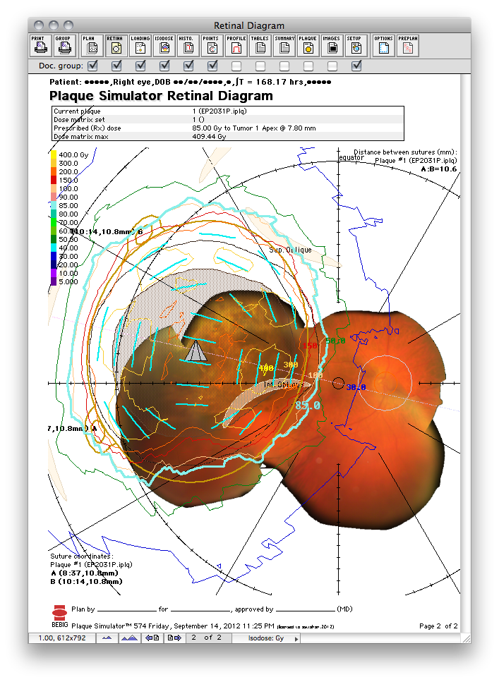
- Page 2, if enabled, can provide an enlarged view of the macular region by reproducing the view created in the retinal diagram window.
- The page advance buttons along the bottom of the window change the page being previewed.
|