OsiriX Tutorial for Plaque Simulator

What is OsiriX?

OsiriX is a DICOM listener and viewer that can manipulate 3-D data sets and export reconstructions of axial, equatorial and sagittal planes which bisect the affected eye as .jpg files. Other reconstructions of interest are the meridian and coronal planes which intersect the tumor apex.

These reconstructions will be imported into Plaque Simulator and used to measure the size and shape of the eye and, in conjunction with ultrasound images and fundus photos, to precisely determine the tumor size, shape and location within the eye.

  • To install OsiriX:
  • Download OsiriX and install on your MacOSX computer.
  • Launch OsiriX by clicking on the Eye of Horus icon on the dock or from the OSX Applications folder.

T-MeridianPlaneFusion.png

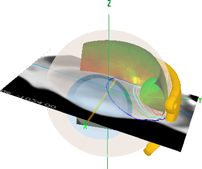
Example of Plaque Simulator fusion of 3D model of eye and plaque with fundus photo and meridian CT reconstruction through the tumor apex with isodose lines.


These are the planar reconstructions for Plaque Simulator that you will create using OsiriX
AxialPlane.png

Axial meridian plane bisecting the eye and intersecting the optic disc (3 to 9 o'clock meridian on the retinal diagram).

EquatorialPlane.png

Equatorial plane bisecting the eye.

SagittalPlane.png

Sagittal meridian plane bisecting the eye (12 to 6 o'clock meridian on the retinal diagram)

T-MeridianPlane.png

Meridian plane bisecting the eye through the tumor apex.

T-CoronalPlane.png

Coronal plane intersecting the tumor apex.


Steps for preparing reconstructions for Plaque Simulator
  • Osirix is a comprehensive MacOSX DICOM listener client that can receive images over your local network. See the OsiriX documentation for assigning an AETitle and other DICOM networking issues. In this tutorial, however, we will assume you are working with a set of transaxial CT images that cover the orbit at <= 3 mm spacing, and that the images were received on portable media such as a CD, DVD or USB memory stick.
  • Insert the CD disk containing the patient's DICOM images into the optical drive of your MacOSX computer.
  • OsiriX will find and load the images from portable media. If OsiriX doesn't automatically find the images, open the CD (or portable device) from the MacOSX Finder and drag the folder containing the images onto OsiriX or its icon.
  • The loaded image sets will appear on the left pane of the OsiriX window.
  • Double-click on the desired study: e.g. Axial slices with 2 mm resolution.
  • A new window will appear.
  • Click on the 2D/3D button at the top of the window.
  • Select the 3D Curved-MPR (Multi Planar Reconstruction) menu item.
  • Adjust the three orthogonal views to align with the appropriate eye.
  • Export axial, coronal, sagittal, tumor-coronal and tumor-meridian reconstructions as .jpg files for subsequent importation into Plaque Simulator.

Launching OsiriX: The initial screen
  • The thumbnail images beneath the patient list represent the available studies for the selected patient.
  • Double-click the thumbnail image of the desired study.
  • The selected study will appear in its own separate window and can be scrolled through the image set.
OsiriXSelection.png

OsiriX: Opened study
OsiriXStudyOpened.png
  • Navigate through the opened study using the horizontal slider located just below the title bar (EYE FOR EYE PLAQUE-KW) of the window that displays the imgages.

OsiriX: Controls and buttons
  • In the tools panel at the top of the screen, click the cursor on the button labeled 2D/3D. It has a gear shaped icon. The 2D/3D dropdown menu will appear.
  • Select the 3D Curved - MPR menu item, the second item on the list.
  • A new window with 4 panes will appear showing three orthogonal reconstructions and a blank pane.
  • Quadrant I: Coronal reconstruction in the upper-right pane.
  • Quadrant II: Sagittal reconstruction in the upper-left pane.
  • Quadrant III: Axial reconstruction in the lower-left pane.
  • Quadrant IV: The lower-right pane is empty.
OsiriX2D3Dmenu.png


OsiriX: The 4-pane window
  • The image manipulation tools are on the left side of the header section.
  • Select a tool from this group in the header by clicking on the desired tool button.
  • OsiriXWindowLevel.png

    Adjust window & level to maximize contrast of the sclera and tumor.

  • Use the translation tool to move the image and the axes crosshairs.
  • OsiriXTranslate.png
    • Placing the cursor over the intersection of the axes crosshairs in any image will produce a cursor that translates the two axes in unison. Drag the crosshairs to the center of the affected eye.
    • Placing the cursor over an axis away from the intersection will produce a rotation-cursor.
    • Drag and rotate the axes until that the yellow axis marks the sagittal meridian (12 to 6 o'clock meridian on the retinal diagram) plane that bisects the eye, the blue axis marks the equatorial plane of the eye, and the purple axis marks the axial meridian plane that bisects the eye through the optic disc (3 to 9 o'clock meridian on the retinal diagram).
    • In the axial image, the yellow axis is aligned with Plaque Simulator's X axis of the eye and passes through the apex of the cornea, the midpoint of the lens, and the posterior pole of the eye.
    • Note that because the patient may not have been looking straight ahead during the scan, the axial, equatorial and sagittal reconstructions of the eye might not be parallel to the axial, coronal and sagittal planes of the body.
OsiriX4Pane.png

OsiriX: Preparing axial, equatorial and sagittal reconstructions for export to Plaque Simulator
  • Magnify to taste.
  • OsiriXmagnify.png

    Use the magnifier tool to adjust the CT image size so that the eye occupies a substantial portion of the pane, but is not obscured by text overlays.

  • For best results with PS, the axial, coronal and sagittal reconstruction panes should be in the range 600 to 800 pixels wide.
  • When the reconstructions are ready, click on the axial pane (lower left quadrant) to select it.
  • A colored BORDER will appear to indicate the selected pane.
  • Click on the File menu > Export > Export to JPEG
  • Name the file appropriately (e.g. 'Axial.jpg' for the axial image).
  • Navigate the file save dialog to find the folder you previously created for the patient (e.g. somewhere in Documents:Plaque Simulator Patients:).
  • Accept the default .jpg settings and save the file in the patient's folder.
  • Save the coronal and sagittal images in the same way. The axial meridian image is required by PS, the sagittal meridian and equatorial planes are optional but useful.
OsiriXOrthogonalPlanes.png

OsiriX: Preparing meridian and coronal reconstructions through the tumor apex for export to Plaque Simulator
  • Rotate the crosshairs in the coronal reconstruction pane to create a meridian reconstruction that intersects the tumor apex. The meridian reconstruction will appear in either the the upper left or lower left quadrant depending upon how far you rotate the crosshairs in the coronal pane. In this example it is the lower left quadrant.
  • In the meridian reconstruction (e.g. lower left quadrant), translate the blue line away from the equator to create a coronal reconstruction (upper right quadrant) that also intersects the tumor apex.
  • Export as .jpg files in the same way as the axial, equatorial and sagittal images. Name the meridian reconstruction 'T-Meridian.jpg'. Name the coronal reconstruction 'T-Coronal.jpg'.
OsirixTumorApexPlanes.png

CT Tutorial | Image Window | Guide Contents