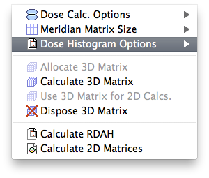
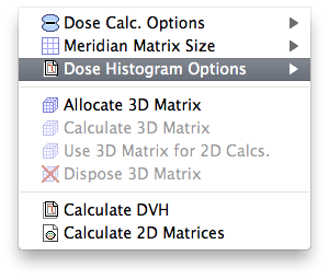
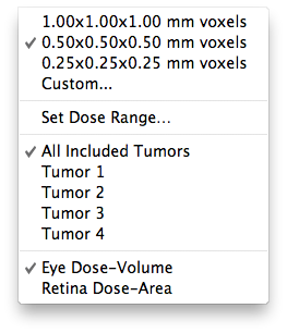
The Dose Histogram Options menu controls the resolution and appearance of dose-histogram calculations.
Retina Dose-Area - set by default, this is the most useful dose histogram because, from a radiobiological perspective, the retina is the most critical region of the eye.
For the retina dose-area calculation (RDAH), a retinal diagram is imagined to be subdivided into pixels. The average dose per pixel is estimated as the dose delivered to a point at the center of each pixel. Calculation time is inversely proportional to the number of pixels.
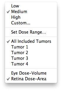
- Low - fastest to calculate, 256 pixel diameter retinal diagram (≈51472 calculation points).
- Medium - a good compromise for routine use, 512 pixel diameter retinal diagram (≈205887 calculation points)
- High - slowest to calculate, 1024 pixel diameter retinal diagram (≈823549 calculation points)
- Custom... - opens a dialog to specify the diameter of the retinal diagram in pixels.
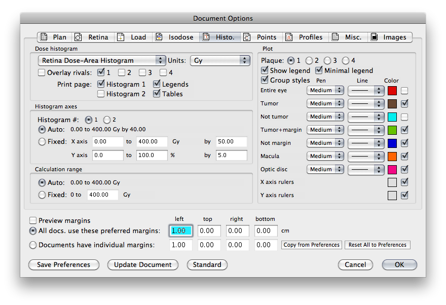
- Set Dose range... - The RDAH calculation and plotting parameters are set in the histogram pane of the Document Options dialog.
- All Included Tumors - the RDAH tumor region includes the active tumor, or, in the case of multiple tumors, all included tumors.
- Tumor 1 - the RDAH tumor region is the base of tumor #1, even if it is not active or included. This enables unused tumors to be repurposed as retinal regions of interest. Note: If you customize the name of a tumor in the Multiple Tumors dialog, that name will appear here rather than 'Tumor 1'.
- Tumor 2 - the RDAH tumor region is the base of tumor #2, even if it is not active or included. This enables unused tumors to be repurposed as retinal regions of interest.
- Tumor 3 - the RDAH tumor region is the base of tumor #3, even if it is not active or included. This enables unused tumors to be repurposed as retinal regions of interest.
- Tumor 4 - the RDAH tumor region is the base of tumor #4, even if it is not active or included. This enables unused tumors to be repurposed as retinal regions of interest.
- Eye Dose-Volume - select to change to dose-volume histogram mode.
- Retina Dose-Area - select to change to dose-area histogram mode.
Eye Dose-Volume - optional, but not very useful, because most of the eye volume is radiobiologically inert vitreous humour.
For the dose-volume calculation, the volume of the eye and tumor are subdivided into voxels. The average dose per voxel is estimated as the dose delivered to a point at the center of each voxel. Calculation time is inversely proportional to voxel size.
- 1.00x1.00x1.00 mm Voxels - fastest to calculate.
- 0.50x0.50x0.50 mm Voxels - a good compromise for the entire eye.
- 0.25x0.25x0.25 mm Voxels - best for tumor volume.
- Custom... - opens a dialog to specify the edge dimension of a cubic voxel.
- Set Dose range... - The DVH calculation and plotting parameters are set in the histogram pane of the Document Options dialog.
- All Included Tumors - the DVH tumor volume includes the active tumor, or, in the case of multiple tumors, all included tumors.
- Tumor 1 - the DVH tumor volume is tumor #1, even if it is not active or included. This enables unused tumors to be repurposed as volumes of interest.
- Tumor 2 - the DVH tumor volume is tumor #2, even if it is not active or included. This enables unused tumors to be repurposed as volumes of interest.
- Tumor 3 - the DVH tumor volume is tumor #3, even if it is not active or included. This enables unused tumors to be repurposed as volumes of interest.
- Tumor 4 - the DVH tumor volume is tumor #4, even if it is not active or included. This enables unused tumors to be repurposed as volumes of interest.
- Eye Dose-Volume - select to change to dose-volume histogram mode.
- Retina Dose-Area - select to change to dose-area histogram mode.