The Eye Physics Model 917

This is Eye Physics' first product, the model EP917 plaque. It is shown fully loaded with 17 seeds. The naming nomenclature differs from subsequent Eye Physics plaques in which the leftmost digits of the plaque name indicate its nominal diameter. In this special case the model name begins with 9 for nostalgic reasons to indicate that the plaque is a descendant of the versatile USC#9 plaque which saw service at the University of Southern California (USC) from the 1980s through 2007. The #9 was the most popular plaque for small and medium size tumors at USC during that era, with several variants including one designed to deliver concurrent hyperthermia. The 17 in the Eye Physics name refers to the number of seed slots.

The EP917 is approximately 1.5 mm thick, it is designed to fit an eye of approximately 25 mm equatorial diameter. It has 17 wells that were manually carved into the plaque's face (ie the concave surface). The openings of these wells (aka slots) are approximately 1.2 mm wide by 5 mm long by 0.8 mm deep. When a seed is mounted in one of these slots it is oriented so that its long axis is approximately tangent to the sphere defined by the plaque face and its upper surface about even with the surface of the plaque face at the seed's center. The slots are therefore slightly deeper at their far ends than at their centers, and their openings in the plaque face are slightly curved.

Loctite4014Big
EP917loaded

The model EP917 is a 2nd generation, manually prototyped plaque intended for small to medium size tumors with GTV base dimensions ≤ 8x10 mm and a 2mm PTV margin at the inner sclera and height ≤ 6 mm measured from the inner surface of the sclera. The plaque has 2 suture eyelets located along its semi-elliptical anterior edge and a broad posterior notch with angled slots to facilitate treatment of small peripapillary tumors. It is cast from 18K standard gold alloy supplied by the David H. Fell Company of Los Angeles, California.

The radionuclide sources (aka seeds) are mounted in the slots using a thin layer of a cyanoacrylate adhesive that is heat tolerant and approved by the FDA for medical instruments. You can learn more about cyanoacrylate glues at this link Tech Tips: Good to Know About Superglue. In general, cyanoacrylate glue hardens very quickly when trapped between two surfaces. The reaction is caused by the condensed water vapour on the surfaces (namely the hydroxyl ions in water). The water comes from the surrounding air. The curing reaction starts at the surface of the bonded material and develops towards the center of the bond. Because of this, thick seams or large blobs of glue may harden less satisfactorily than surface-to-surface bonds with good fit. In a thick blob of glue, a polymerisation reaction may stop before it reaches the centre of the blob. A rule of thumb is that seams thicker than 0.25 mm should be avoided.

Hence, with cyanoacylate, a thin layer between conformal surfaces provides the strongest bond. Seeds bonded by cyanoacrylate can be gently removed by soaking the plaque in an acetone bath until the glue dissolves. The plaques should then be cleaned down to bare metal by brushing the seed slots with a toothbrush followed by ultrasonic cleaning in acetone.


History of the USC#9 and EP917

USC9FirstGenThe USC#9 plaque originated in the late 1980s as this very primitive prototype. It was conceived as a geometrically more reproducible alternative to manually fabricated designs of that era such as this plaque from Philadelphia which consisted of seeds glued to the face of a plastic shell backed with a lead foil.Philadelpia100x90 The USC#9 would be cast from gold rather than use hazardous lead for posterior shielding and it would use cyanoacrylate to mount the seeds rather than dental acrylic, making it much easier to recover the seeds.

Both of these plaques were dosimetrically minimalist designs in the sense that they made no attempt to shield the patient from any forward or laterally directed radiation emitted from the plaque, and unlike the alternative COMS designs, these plaques were thinner and the seeds not embedded in a costly single use silicone carrier. The dominant absorption mechanism for low energy I-125 radiation is the photoelectric effect for which the COMS silicone carrier is significantly more attenuating than water owing to the higher effective atomic number (compared to water) of the carrier material which is mostly silicon. This inhomogeneity surrounding the seeds adds complexity to dosimetry calculations. The minimalist nature of these early plaques was all well and good because brachytherapy planning systems in the 1980s could not readily account for seed collimation and other inhomogeneities on such a small physical scale.

USC9smallIn 1993 the USC#9 design was refined and polished, the crude rectangular gluing slots were manually rounded to better match the cylindrical shape of I-125 seeds in order to provide a more conformal surface-to-surface fit for cyanoacrylate bonding of the seeds to the plaque face. The depth of the slots of the 1993 revision were less than half the diameter of an I-125 seed, preserving the plaque's dosimetrically minimalist design. Slot collimation calculations, introduced around 1997, are not applicable to the USC#9. The 1993 version was also cast a bit thinner than the 1989 model in order to make it lighter and easier to implant.

USC9_90x80The 1993 casting of the USC #9 plaque predates the incorporation of Eye Physics, LLC by more than a decade. However, a few plaques of this design were purchased via direct arrangement between the jeweler who manufactured the plaques for USC and various interested parties.
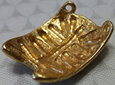
USC9_1stGen162x120In the mid 1990s Astrahan used a Dremel tool to experimentally modify a 1989 casting of the USC#9 with deeper slots to provide an even more secure cyanoacrylate bonding surface and observed that the modification also resulted in some collimation of laterally directed radiation emitted from the plaque. This is the home-made experimental model that eventually became a workhorse at USC and inspired the research effort that led to the plaque described by Astrahan et al. in 1997 and a decade later inspired the EP917.
EP917CarvedPrototype

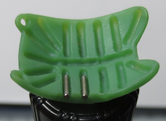
All Eye Physics plaques begin as wax, resin or brass prototypes from which molds are created. The final gold castings are made from these molds. This is a wax EP917 prototype with a section carved away to illustrate how seeds reside in the slots.

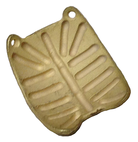
Below is an EP917 that has just come from casting. After casting, the residual plaster dust is removed in a tumbler and the plaque is hand polished to achieve its final appearance.
EP917BeforePolishing286x294

In 2007 Eye Physics acquired the distribution rights for the plaques produced by the jeweler for USC and commissioned a new casting of the USC#9 pattern with deeper slots. Decayed seeds were used to manually test the shape and depth of the slots. The 2007 revision of the USC#9 for Eye Physics was designated as the model EP917.


These are the molds that have been used since 1989 to cast the USC#9 and EP917 plaques.

USC9History

The EP917 vs Astrahan's 1997 plaque

Eye Physics, LLC was incorporated in 2007 to continue the development of the plaque simulation software begun at USC in the late 1980s and described by Astrahan et al., An interactive treatment planning system for ophthalmic plaque radiotherapy, International Journal of Radiation Oncology, Biology, Physics, 18: 679-687, 1990 and later by Astrahan et al. Conformal Episcleral Plaque Therapy, International Journal of Radiation Oncology, Biology, Physics, 39: 505-519, 1997 and numerous other references.

The one-off research plaque, pictured on the left side of Fig.3 from the 1997 article, had little in common with either the USC#9 or the COMS style plaques. It had 25 deep collimating slots, no seed carrier, no posterior notch, and the anterior end is not elliptical. The size, shape and slot design of the 1997 device were inspired by lessons learned from research efforts to create computer models of the USC#9 and COMS plaques, but the 1997 device is clearly very different from a COMS plaque or a USC#9 or its subsequent descendent the EP917. The plaque cast for the 1997 publication had no specific manufacturing connection to the USC#9 plaque of that era other than it used the same gold alloy and was cast by the same jeweler.

SlottedPlaque1997

The process used to fabricate and prototype the deep, sharply angled, well defined seed slots of the 1997 plaque was developed by Astrahan and is completely different from methods used to prototype the USC#9 and EP917. The 1997 method was very costly and has been superseded by the very affordable digital stereolithography 3D printing technology developed by Astrahan that is now used by Eye Physics for its newest plaques.

The shape and dimensions of the 1997 plaque and its slots were selected for treating medium to tall tumors, whereas the EP917 is intended for small to medium height tumors. All of the slots in the 1997 plaque used the same slot design.

In the current digitally prototyped plaques, there is a wide variation in slot dimensions within a plaque based on the dosimetric objectives of the plaque. For example, the slots of most 3rd generation Eye Physics plaques become progressively shallower as one moves radially outward from the center towards the perimeter of the plaque, effectively increasing the apparent source strength of peripheral seeds without having to use seeds of different activities. This approach emulates the classic Paterson-Parker brachytherapy system which was designed to create uniform planar implants using a nonuniform distribution of source strength which was more heavily weighted at the periphery of the implant.

To clarify for any persons that may be confused, Eye Physics has never stated, advertised or implied in any way whatsoever that the EP917 uses any of the Astrahan et al. 1997 or current 3rd generation digital slot fabrication technology. All references to the EP917 have always stated that the EP917 is a 2nd generation manually prototyped device.

EP917vs1997

How to distinguish between the USC#9 and the EP917

In the color picture on the right, the left side is a wax prototype of the USC#9 (1993 revision) plaque. In 1993 the USC#9 seed wells were revised to provide a more conformal fit to the seeds in order to strengthen cyanoacrylate bonding compared to the primitive flat bottomed indentations of the original USC#9 of the 1980s shown below.

USC9FirstGen The USC#9 from 1989.

The center channel of the USC#9 is one continuous groove which permitted up to three I-125 seeds (or a thermocouple array for hyperthermia use) to be placed in the channel. If only one or two seeds were placed in the center channel, their positions could be customized to better fit the tumor dimensions. This design was very flexible but added some uncertainty when mounting the seeds in the plaque to reproduce a treatment plan.

On the right side is a wax prototype of the EP917 (developed for Eye Physics in 2007). The slots of the 917 are deeper than the USC#9, the goal was to position the tops of the seeds even with the plaque face in order to provide a superior bonding surface for the cyanoacrylate and to provide some collimation of laterally directed radiation. Decayed seeds were used to shape the wells. An edge has been carved away to illustrate how the seeds appear in the slots, their tops even with the plaque face. Center channel uncertainty was addressed in the EP917 by placing small separator bumps in the channel in order to create three distinct seed positions.

USC9vsEP917

USC9Loaded

Above is a fully loaded (17 seeds) 1993 USC#9 wax prototype (gold USC#9s have not been available or manufactured since 2007).

USC9onEye

The top half of the seeds are visible sandwiched between the USC#9 plaque face and this 25 mm diameter glass eye.


EP917loaded

This is a fully loaded (17 seeds) EP917 plaque.

QuarterView

No portion of any seed is visible between the EP917 plaque face and this 25 mm diameter glass eye. The face and perimeter of the plaque are in close contact with the sclera demonstrating that the seed tops are indeed approximately even with, or slightly below the spherical surface of the plaque face.


Modeling the EP917

Plaque Simulator creates a 3D model of a plaque and its shell, suture and seed coordinates from a high resolution photograph of the plaque. Illustrated here is a 3D model of a translucent single I-125 seed mounted in one of the slots of the 3D plaque model viewed face on.

OneSeedLoading
OneSeedSetup

Illustrated above is a 3D rendering of the EP917 model positioned under an equatorial tumor with isodose lines in the coronal plane through the seed axis.

The Plaque Simulator (PS) software uses the TG43 dosimetry formalism with a modifier derived from TLD measurements to approximate the contribution of gold fluorescence. Collimation is estimated by ray-tracing between the dose calculation point and the line source representing a seed and assuming that all primary radiation which encounters the gold plaque shell is fully absorbed in the shell. This is a fast and reasonable first approximation because at I-125 energies most attenuation is photoelectric and the half value layer for I-125 in lead (whose atomic number is close to that of gold) is 0.02 mm.

The slightly curved seed slots of the EP917 are modeled by cutting rectangular openings into the tessellated face (concave surface) of the computer model of the gold shell and then seaming those openings with the surrounding triangular facets that model the face of the plaque. The openings are tangent to the sphere of the face at their centers. The depth of the slots in the EP917 were designed to keep the upper edge of a 0.8 mm diameter seed approximately even with the face of the plaque at the seed center. To accelerate the ray trace calculations, the sides and bottom of a slot are approximated using 10 triangular facets.

Simplifying the geometry at the bottom of a slot in the computer model by using a small number of flat facets to approximate the curved bottom is irrelevant to PS dosimetry because the ray-traced dosimetry engine assumes that all primary radiation emitted from a seed that encounters the sides and bottom of a slot is completely attenuated within the plaque itself. The precise geometry of the slot inside the plaque shell might be of academic interest to a Monte Carlo dosimetry engine that accounts for secondary scatter and fluorescence effects.

EP917Wireframe

When the plaque curvature matches the scleral curvature, the outer surface of a seed's titanium shell will come in contact with the sclera above the seed's center, and will be about 0.15 mm offset from the sclera above the ends of a 3 mm long linear source (plotted in blue) within the seed.

Because the seed axes and slot openings are tangent to the eye at their centers, most dosimetric variation between modeling the slot corners as curved versus approximating the corners as right angles will occur outside of the eye. The shallow slots used in this plaque only provide significant collimation of the eye from an edge of the slot opening that parallels the seed axis and is also very close to the axis as illustrated on the left below.

The axial ends of the slots are too far away and at too shallow an angle from the linear source to provide the eye with much collimation as illustrated below right. Fortunately, the seeds internal self attenuation (anisotropy) is greatest along their axes so the need for collimation in the axial direction is not as critical as one might initially think in terms of sparing healthy tissue adjacent to the plaque.

SlotDetail

The rectangular opening of the collimating slot as specified in the PS software is seamed with the triangular tessellating facets that model the curved face of the plaque to create a curved opening at the plaque face that is a few tenths of a mm longer than the length of the rectangle. For the EP917, PS uses a 4.8 to 4.9 mm slot length which results in an effective collimating length of about 5.7 mm in the curved face as illustrated above. The slot width is nominally 1.2 mm. The effective curved collimating width at the face varies slightly over the length of some of the slots according to the seaming facets, sometimes being slightly wider and rounded near the corners. A proper Monte Carlo geometry to compare with PS will need to also model the curved nature of the seed slot openings and the perimeter lip of the plaque in contact with the sclera.

Collimation

Measuring the EP917

Eye Physics anticipates a variance of dimensional averages of several hundredths of a mm between plaque castings. These dimensional variations are not considered clinically significant. Small variances are a limitation of the lost-wax manufacturing process and are related to differences in the wax castings, alloy cooling/contracting and mold temperature. Eye Physics uses a digital readout micrometer jig with a precision of 0.0001 inch and a custom 0.8 mm diameter hemispherical rounded tip to measure plaque thickness and slot depths. Variances in plaque thickness do not affect dosimetry.

For the EP917 casting shown here, the average shell thickness was measured to be 1.485 mm and the average slot depth was 0.754 mm at the slot centers. Due to curvature of the plaque face, the slots are about 1 mm deep at the long axis corners. PS version 5 modeled the slots as being 0.8 mm deep at their centers. In PS6, which is capable of more precise modeling, the default EP917 model considers the slots to be 0.75 mm deep at their centers with 0.05 mm of the seed casing rising above the curve of the plaque face at the slot centers. The entire plaque face is then offset from the eye by 0.05 mm to keep the seeds in contact with the sclera at their centers. The depth of each slot at its center may optionally be measured and entered into the PS5 and PS6 default EP917 files to create a customized model for a specific casting.

DepthSheet
DigitalMicrometer

Gold thickness is measured between the micrometer anvil and the bottom of a slot at its axial center, and then between the anvil and the slot edges bracketing the center. The ratchet mechanism assures consistent measurement pressure. The difference between the slot bottom and the average of the bracketing edge measurements is the nominal slot depth.

slot11botom
slot11top