
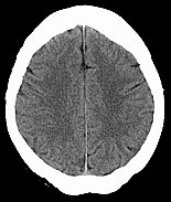
Madena is a medical image viewer and image processing tool. Madena can open all QuickTime compatible image file formats as well as import most Digital Imaging and Communications in Medicine (DICOM) and ACR/NEMA file formats. DICOM is the current industry standard for medical images and is widely used for CT, MR and US images. Madena can open both individual image files or all image files found within a folder and (optionally) all enclosed subfolders in a single command. DICOM images are automatically organized into viewing and processing subsets according to their study and series tags. Quicktime images are grouped into subsets according to the folder in which they were found.
Madena currently supports the following 8-bit and 16-bit DICOM formats: single and multiple frame files, MONOCHROME1, MONOCHROME2, RGB, HSV, PALETTE_COLOR, YBR_FULL, RLE compression, and some encapsulated JPEG (baseline encoding only) files. Madena also imports the ANALYZE 7.5 file format and 16-bit TIFF files. For DICOM and ANALYZE files which contain multiple frames, all the frames can be processed simultaneously or an individual frame can be extracted from the file. Images can be exported to most of the QuickTime supported graphics file formats (eg .jpg, .gif, .pct, etc...) as well as to DICOM and ANALYZE format. Multiple frame images can be exported as QuickTime movies. No additional drivers or licenses are required. Comprehensive "window & level" functionality is provided for 16-bit images. Orthogonal reconstructions of multiplanar (MP) image sets (up to 1024 images per series) at 1 pixel increments are supported. DICOM files can be anonymized individually or in bulk.
 Monochromatic medical images consist of 8-bit (256 levels) or 16-bit (65536 levels) pixels whose values are often referred to as "densities". The term "density" is derived from the original concept of "optical density" (OD) as it is applied to radiological X-Ray films which are viewed using transmitted (as opposed to reflected) light. Optical density is defined as Log(Io/It) where Io and It are the light incident upon, and light transmitted through the film. An OD of 1.0 would mean that 10% of the incident light was transmitted through the film, an OD of 2.0 would mean 1% transmission and so on.
The "density" of a pixel in a modern medical image might represent X-ray attenuation in a patient (eg. a mammogram, a chest x-ray or other radiograph), electron density (CT), proton relaxation time (MRI) or radioisotope distribution in the body (eg. Gallium or PET scans). Medical image densities have historically been displayed as shades of gray. Madena provides powerful viewing options for these images. Images can be enhanced using common linear brightness and contrast functions, as well as non-linear function such as bias and gain. 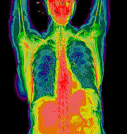 All, or a selected range of densities can be tinted or isodensity colorized. Images can be rescaled, rotated, and translucently fused together (eg. fusion of CT and MRI images, or fusion of a chest x-ray and a gallium scan). Points and regions of interest can be drawn on the images and their areas measured. The images can be calibrated, measured, and printed. Printed images can be enlarged to "life-size" or greater and printed across multiple sheets of paper if necessary.
Madena was originally conceived and coded by Prof. Melvin Astrahan for Dr. Giske Ursin of the University of Southern California School of Medicine to identify and quantify the extent of radiographic density patterns on a mammogram by measuring the percentage of the mammographic image that contains densities in a specified range, and to track changes in density patterns which occur over time or with medical treatment. (Ursin G, Astrahan M, Salane M, Parisky Y, Pearce J, Daniels J, Pike M, Spicer D: The Detection of Changes in Mammographic Densities. Cancer Epidemiol Biomarkers Prev. 1998 Jan;7(1):43-7.)
Madena can be used as a medical image viewer for both monochome and color images. If an image is not natively 16-bit monochrome (eg. an 8-bit or an RGB color image), Madena will automatically create an equivalent 16-bit monochrome dataset as well. You can switch back and forth between the original and the 16-bit monochome version. Most of Madena's image processing functions work with both the original and the 16-bit version, and with multiplanar image sets. Madena was originally conceived as a tool to quickly count the number of pixels in a user defined range of gray levels (or "densities") in a mammogram. To assist in range definition and pattern recognition, a colorful tint can be applied across a range of densities, isodensity bands can be tinted with different colors, isodensity contours plotted, and regions of interest (ROI) defined. For each ROI, Madena calculates the mean gray level and standard deviation, counts the total number of pixels, and the number of tinted pixels, the distribution of pixels in "isodensity" bands, and calculates the area in square cm (and volume in cubic cm for MP sets) if the images are calibrated.
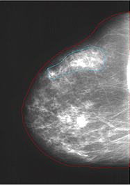
ROI parameters obtained with Madena can be exported to a Microsoft Excel spreadsheet for further analysis. For instance, when analyzing mammograms, a ROI corresponding to the entire breast is first defined. A ROI corresponding to a subregion is then defined. If you want to know the percentage of the breast that falls within a subregion, it can be calculated by dividing the number of pixels in the subregion by the total number of pixels in the entire breast. If you want to know the percentage of the breast that falls within a range of densities, it can be calculated by dividing the number of tinted pixels in the subregion ROI by the total number of pixels in the entire breast ROI. For mammography analysis, specialized training is required to identify where regions of interest should be drawn, and which densities should be tinted on mammograms. We suggest that you conduct the density reading and the outlining of the total breast area in separate steps.
Of course, no mammogram training is required for casual and recreational users of Madena as a general purpose medical imaging tool. Just have fun, and please let us know what you are using Madena for!