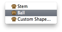
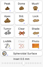
In the Retinal Diagram window's tumor controls group, click the Mushroom button,
 and select Ball from the contextual menu (control-click or right mouse button click the mushroom button to display the contextual menu) to give the tumor a ball or button-like shape.